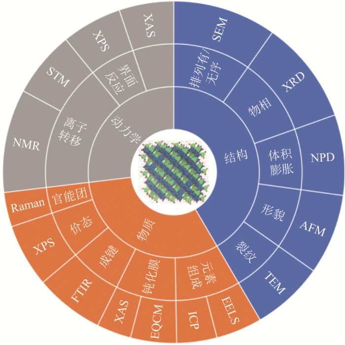
The increasing demand for energy storage requires energy storage devices to have greater capacity, and there are high hopes for lithium-ion batteries (LIBs) in the energy storage field. Their structural stability as cathode materials and their voltage profiles for insertion/extraction directly determine the specific energy and power densities of the battery system. In recent years, research related to these characteristics has remained the core issue in the LIB research field, particularly the characterization of the material structure and electrochemical behavior. Important real-time and in-situ strategies were employed in designing and developing more types of materials with excellent performance. For the cathode materials, detailed insights, such as their microstructure, chemical composition, ion valence and states, character of morphology, ion transport, and electron transfer, are beneficial in the preparation, structure design and modification of electrode materials. In this review, the operating principles, usage scenarios, and corresponding information of characterization techniques are introduced, and some examples that use these techniques to characterize LIB anode materials are listed. Finally, the advantages and disadvantages of current characterization techniques are compared, and the major challenges in research studies are discussed. Hence, this article summarizes the typically used technologies that are applied in monitoring the structural changes and surface-interface behaviors of cathode materials, including the microscopic imaging, phase analysis, composition and chemical valence, and bonding and functional groups to provide a reference for the combined utilization of various characterization technologies and to promote the development of an ideal electrode material.
MU Yue. Methods of investigating structural evolution and interface behavior in cathode materials for Li-ion batteries[J]. Energy Storage Science and Technology, 2021, 10(1): 7-26
Fig.2
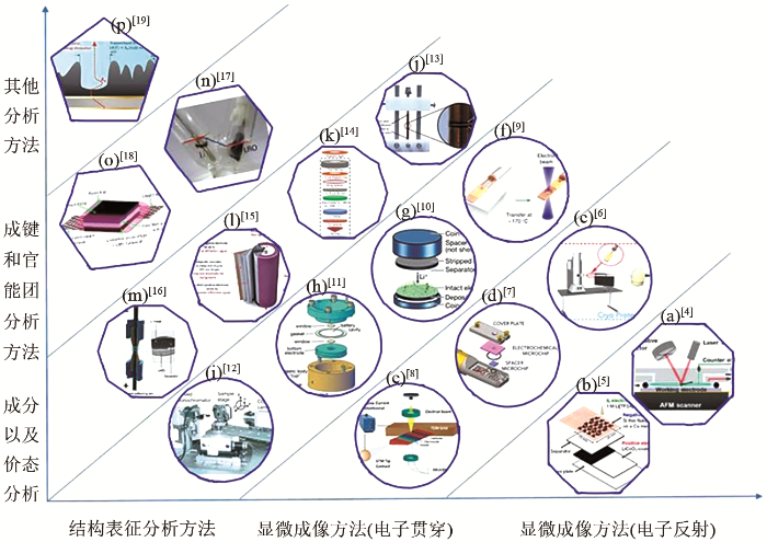
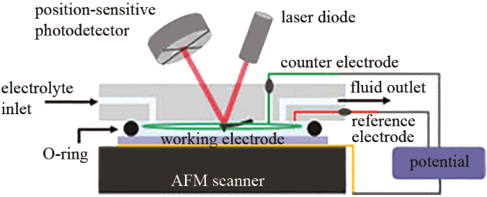
Some cell setups used for in situ characterization[4-19]: (a) for AFM; (b), (c)for SEM; (d) for TEM; (e) for STEM; (f), (g) for Cryo-EM; (h), (i), (j) for X-ray characterization; (k), (l) for neutron characterization; (m) for XRD-CT; (n) for FTIR; (p) for EQCM
Fig.3
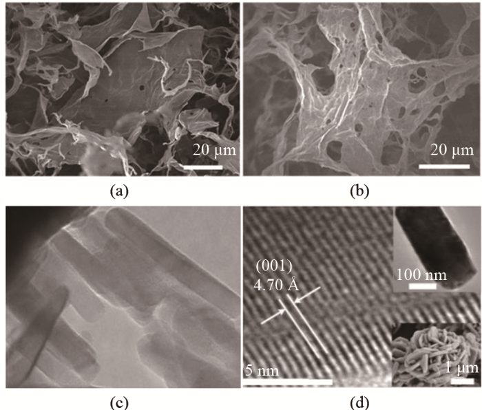
SEM images of GO, weight ratio of balls to GO aqueous suspension: (a) 0.6,; (b) 1.2[30]; (c) TEM image of LiMnPO4[31]; (d) HRTEM image of NCM-H[38]
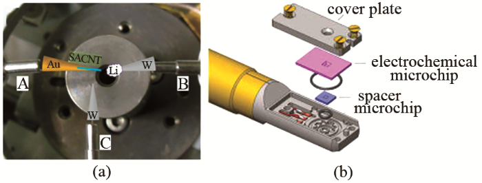
SEM和TEM可以使用环境腔技术(environmental scanning electron microscopy,ESEM)实现原位观察。而原位观察需要在电池运行过程中使探测电子束进入电池体系,因此在电池设计上和传统电池差别很大。这一类技术目前没有在正极表征上进行使用,但在负极的研究中已经开始使用,例如,Zheng等[39]设计了一种原位电池,如图所示使用环境腔技术,然后Au针尖上固定直壁碳纳米管做正极,W针尖上固定Li做负极,另一W针尖用以划擦Li的表面,保证原始Li暴露出来,使用这个原位装置观察充放电循环时的结构演变。后续在正极材料,尤其是单晶结构正极材料的表面行为表征方面将会有潜在的应用前景。Unocic等[7]使用Poseiden 500原位微流体电化学S/TEM表征系统,如图4所示,此系统本质上是一个密封在TEM支架端的三电极微流体电化学电池。该支架集成了微流体输送系统和电气触点,能控制电解液的输送并进行实时的电化学测量。制作的微型器件以玻璃碳和铂微带为电极,并以SiNx作为观察窗口。
Fig.5
(a) HADDF-STEM image of LMR, showing structure of defect spinel structure with empty 16c octahedral sites[32]; (b) ABF-STEM image of processed Li2MnO3[33]; (c) AFM images of thin-film V2O5 cathodes on different states[34]
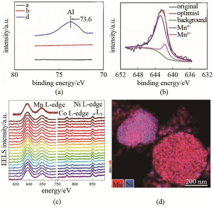
Fig.12
(a) XPS spectra of 3% LiAlO2 doped sample of Mn element; (b) is XPS spectra of sample in (a) after 100 cycles[67]; (c) Transition metal L-edge EELS spectra along scanning pathway[59]; (d) EDX image of Ti-doped LiNi0.5Mn1.5O4-δ[60]
Fig.15
(a) FTIR patterns of LRO electrode[17]; (b) Raman spectra of pristine and cycled Li1.2Ni0.16Mn0.56Co0.08O2[69]; (c) NMR shows effect of aluminum content on Al local structure of NCM 523[70]
NMR是使用频率为MHz级别的电磁波照射分子,使磁性原子核在外磁场中发生磁能级的共振跃迁,并获得吸收信号,得出一个射频辐射吸收的光谱。获得NMR信号需要核子数为奇数的原子。NMR具有高的能量和空间的分辨能力。此外基于NMR的核磁共振成像(NMRI)也在分析电池电极时使用[72]。NMR中的魔角旋转核磁共振谱(MAS NMR)可以定性、定量地表征Al的配位信息,分析其在晶格中的作用,这对NCA正极材料的研究来说很有意义。Dogan等[70]就使用27Al MAS NMR直接观察NCA正极材料中的Al晶格环境[图15(c)],得知对NCA而言,晶体中的Al更倾向于和6个Ni配位,成蜂巢状,而增加Co的含量会增加铝的位错。而Al含量增大会使铝酸盐增多,出现偏析倾向,同时分析得知NCA中Al的在晶格中的最大占有率约为5%。Yim等[71]使用硼酸三苯作为添加剂改善CEI性能,使用原位NMR分析硼酸三苯对残锂清除的影响,硼酸三苯参与反应的CEI层抑制了电解液的分解,从而增强了电极表面的稳定性。
YAN Pengfei, ZHENG Jianming, GU Meng, et al. Intragranular cracking as a critical barrier for high-voltage usage of layer-structured cathode for lithium-ion batteries[J]. Nature Communications, 2017, 8(1): doi: 10.1038/ncomms14101.
PANG W K, LIN H, PETERSON V K, et al. Effects of fluorine and chromium doping on the performance of lithium-rich Li1+xMO2 (M=Ni, Mn, Co) positive electrodes[J]. Chemistry of Materials, 2017, 29(24): 10299-10311.
ZENG Y, CHIU H, RASOOL M, et al. Hydrothermal crystallization of Pmn21 Li2FeSiO4 hollow mesocrystals for Li-ion cathode application[J]. Chemical Engineering Journal, 2019, 359: 1592-1602.
LIU Xingrui, WANG Dong, WAN Lijun. Progress of electrode/electrolyte interfacial investigation of Li-ion batteries via in situ scanning probe microscopy[J]. Science Bulletin, 2015, 60(9): 839-849.
CHEN C Y, SANO T, TSUDA T, et al. In situ scanning electron microscopy of silicon anode reactions in lithium-ion batteries during charge/discharge processes[J]. Scientific Reports, 2016, 6: doi: 10.1038/srep36153.
WANG X F, LI Y J, MENG Y S. Cryogenic electron microscopy for characterizing and diagnosing batteries[J]. Joule, 2018, 2(11): 2225-2234.
UNOCIC R R, SACCI R L, BROWN G M, et al. Quantitative electrochemical measurements using in situ ec-S/TEM devices[J]. Microscopy and Microanalysis, 2014, 20(2): 452-461.
WANG Z Y, SANTHANAGOPALAN D, ZHANG W, et al. In situ STEM-EELS observation of nanoscale interfacial phenomena in all-solid-state batteries[J]. Nano Letters, 2016, 16(6): 3760-3767.
LI Y Z, LI Y B, PEI A, et al. Atomic structure of sensitive battery materials and interfaces revealed by cryo-electron microscopy[J]. Science, 2017, 358(6362): 506-510.
ZACHMAN M J, TU Z, CHOUDHURY S, et al. Cryo-STEM mapping of solid-liquid interfaces and dendrites in lithium-metal batteries[J]. Nature, 2018, 560(7718): 345-349.
BORKIEWICZ O J, SHYAM B, WIADEREK K M, et al. The AMPIX electrochemical cell: A versatile apparatus for in situ X-ray scattering and spectroscopic measurements[J]. Journal of Applied Crystallography, 2012, 45(6): 1261-1269.
LYU Yingchun, LIU Yali, CHENG Tao, et al. High-throughput characterization methods for lithium batteries[J]. Journal of Materiomics, 2017, 3(3): 221-229.
LIU Hao, ALLAN P K, BORKIEWICZ O J, et al. A radially accessible tubular in situ X-ray cell for spatially resolved operando scattering and spectroscopic studies of electrochemical energy storage devices[J]. Journal of Applied Crystallography, 2016, 49(5): 1665-1673.
LIU D X, WANG J H, PAN K, et al. In situ quantification and visualization of lithium transport with neutrons[J]. Angew Chem Int Ed Engl, 2014, 53(36): 9498-9502.
BOULET-ROBLIN L, SHEPTYAKOV D, BOREL P, et al. Crystal structure evolution viaoperando neutron diffraction during long-term cycling of a customized 5 V full Li-ion cylindrical cell LiNi0.5Mn1.5O4vs. graphite[J]. Journal of Materials Chemistry A, 2017, 5(48): 25574-25582.
FINEGAN D P, VAMVAKEROS A, TAN Chun, et al. Spatial quantification of dynamic inter and intra particle crystallographic heterogeneities within lithium ion electrodes[J]. Nature Communications, 2020, 11(1): doi: 10.1038/s41467-020-14467-x.
TU Wenqiang, XIA Pan, ZHENG Xiongwen, et al. Insight into the interaction between layered lithium-rich oxide and additive-containing electrolyte[J]. Journal of Power Sources, 2017, 341: 348-356.
KEY B, BHATTACHARYYA R, MORCRETTE M, et al. Real-time NMR investigations of structural changes in silicon electrodes for lithium-ion batteries[J]. Journal of the American Chemical Society, 2009, 131(26): 9239-9249.
SHPIGEL N, LEVI M D, SIGALOV S, et al. In situ hydrodynamic spectroscopy for structure characterization of porous energy storage electrodes[J]. Nature Materials, 2016, 15(5): 570-575.
BRANT W R, LI D, GU Q F, et al. Comparative analysis of ex-situ and operando X-ray diffraction experiments for lithium insertion materials[J]. Journal of Power Sources, 2016, 302: 126-134.
GANAPATHY S, ADAMS B D, STENOU G, et al. Nature of Li2O2 oxidation in a Li-O2 battery revealed by operando X-ray diffraction[J]. Journal of the American Chemical Society, 2014, 136(46): 16335-16344.
BORKIEWICZ O J, WIADEREK K M, CHUPAS P J, et al. Best practices for operando battery experiments: Influences of X-ray experiment design on observed electrochemical reactivity[J]. The Journal of Physical Chemistry Letters, 2015, 6(11): 2081-2085.
GU M, PARENT L R, MEHDI B L, et al. Demonstration of an electrochemical liquid cell for operando transmission electron microscopy observation of the lithiation/delithiation behavior of Si nanowire battery anodes[J]. Nano Letters, 2013, 13(12): 6106-6112.
HOLTZ M E, YU Y, GUNCELER D, et al. Nanoscale imaging of lithium ion distribution during in situ operation of battery electrode and electrolyte[J]. Nano Letters, 2014, 14(3): 1453-1459.
CHEN D C, MAHMOUD M A, WANG J, et al. Operando investigation into dynamic evolution of cathode-electrolyte interfaces in a Li-ion battery[J]. Nano Letters, 2019, 19(3): 2037-2043.
MA Kaijie, ZHANG Yunong, LIU Le, et al. In situ mapping of activity distribution and oxygen evolution reaction in vanadium flow batteries[J]. Nature Communications, 2019, 10(1): doi: 10.1038/s41467-019-13147-9.
LIN Ruoqian, HU Enyuan, LIU Mingjie, et al. Anomalous metal segregation in lithium-rich material provides design rules for stable cathode in lithium-ion battery[J]. Nature Communications, 2019, 10(1): doi: 10.1038/s41467-019-09248-0.
ZHANG Jienan, LI Qinghao, OUYANG Chuying, et al. Trace doping of multiple elements enables stable battery cycling of LiCoO2 at 4.6 V[J]. Nature Energy, 2019, 4(7): 594-603.
XIE Song, REN Lixiang, YANG Xiaoyong, et al. Influence of cycling aging and ambient pressure on the thermal safety features of lithium-ion battery[J]. Journal of Power Sources, 2020, 448: doi: 10.1016/j.jpowsour.2019.227425.
DING D, MAEYOSHI Y, KUBOTA M, et al. Holey reduced graphene oxide/carbon nanotube/LiMn0.7Fe0.3PO4 composite cathode for high-performance lithium batteries[J]. Journal of Power Sources, 2020, 449: doi: 10.1016/j.jpowsour.2019.227553.
WANG D, BUQA H, CROUZET M, et al. High-performance, nano-structured LiMnPO4 synthesized via a polyol method[J]. Journal of Power Sources, 2009, 189(1): 624-628.
ZHENG Jianming, XU Pinghong, GU Meng, et al. Structural and chemical evolution of Li- and Mn-rich layered cathode material[J]. Chemistry of Materials, 2015, 27(4): 1381-1390.
QING Renpeng, SHI Jile, XIAO Dongdong, et al. Enhancing the kinetics of Li-rich cathode materials through the pinning effects of gradient surface Na+ doping[J]. Advanced Energy Materials, 2016, 6(6): doi: 10.1002/aenm.201501914.
COHEN Y S, AURBACH D. Surface films phenomena on vanadium-pentoxide cathodes for Li and Li-ion batteries: In situ AFM imaging[J]. Electrochemistry Communications, 2004, 6(6): 536-542.
ALVARADO J, SCHROEDER M A, ZHANG Minghao, et al. A carbonate-free, sulfone-based electrolyte for high-voltage Li-ion batteries[J]. Materials Today, 2018, 21(4): 341-353.
LI Xinlu, KANG Feiyu, BAI Xinde, et al. A novel network composite cathode of LiFePO4/multiwalled carbon nanotubes with high rate capability for lithium ion batteries[J]. Electrochemistry Communications, 2007, 9(4): 663-666.
DOHERTY C M, CARUSO R A, SMARSLY B M, et al. Hierarchically porous monolithic LiFePO4/carbon composite electrode materials for high power lithium ion batteries[J]. Chemistry of Materials, 2009, 21(21): 5300-5306.
ZHENG Hao, XIAO Dongdong, LI Xing, et al. New insight in understanding oxygen reduction and evolution in solid-state lithium-oxygen batteries using an in situ environmental scanning electron microscope[J]. Nano Letters, 2014, 14(8): 4245-4249.
PENNYCOOK S J, BOATNER L A. Chemically sensitive structure-imaging with a scanning transmission electron microscope[J]. Nature, 1988, 336(6199): 565-567.
OKUNISHI E, ISHIKAWA I, SAWADA H, et al. Visualization of light elements at ultrahigh resolution by STEM annular bright field microscopy[J]. Microscopy and Microanalysis, 2009, 15(S2): 164-165.
HU Enyuan, YU Xiqian, LIN Ruoqian, et al. Evolution of redox couples in Li- and Mn-rich cathode materials and mitigation of voltage fade by reducing oxygen release[J]. Nature Energy, 2018, 3(8): 690-698.
INABA M, TOMIYASU H, TASAKA A, et al. Atomic force microscopy study on the stability of a surface film formed on a graphite negative electrode at elevated temperatures[J]. Langmuir, 2004, 20(4): 1348-1355.
JAISER S, KUMBERG J, KLAVER J, et al. Microstructure formation of lithium-ion battery electrodes during drying—An ex-situ study using cryogenic broad ion beam slope-cutting and scanning electron microscopy (Cryo-BIB-SEM)[J]. Journal of Power Sources, 2017, 345: 97-107.
WANG Xuefeng, ZHANG Minghao, ALVARADO J, et al. New insights on the structure of electrochemically deposited lithium metal and its solid electrolyte interphases via cryogenic TEM[J]. Nano Letters, 2017, 17(12): 7606-7612.
LI Y Z, HUANG W, LI Y B, et al. Correlating Structure and function of battery interphases at atomic resolution using cryoelectron microscopy[J]. Joule, 2018, 2(10): 2167-2177.
SCHIPPER F, DIXIT M, KOVACHEVA D, et al. Stabilizing nickel-rich layered cathode materials by a high-charge cation doping strategy: Zirconium-doped LiNi0.6Co0.2Mn0.2O2[J]. Journal of Materials Chemistry A, 2016, 4(41): 16073-16084.
RYU H H, PARK K J, YOON C S, et al. Capacity fading of Ni-rich Li [NixCoyMn1–x–y]O2 (0.6≤x≤0.95) cathodes for high-energy-density lithium-ion batteries: bulk or surface degradation?[J]. Chemistry of Materials, 2018, 30(3): 1155-1163.
GOONETILLEKE D, SHARMA N, PANG W K, et al. Structural evolution and high-voltage structural stability of Li(NixMnyCoz)O2 electrodes[J]. Chemistry of Materials, 2018, 31(2): 376-386.
MOHANTY D, DAHLBERG K, KING D M, et al. Modification of Ni-rich FCG NMC and NCA cathodes by atomic layer deposition: Preventing surface phase transitions for high-voltage lithium-ion batteries[J]. Scientific Reports, 2016, 6(1): doi: 10.1038/srep26532.
KLEINER K, STREHLE B, BAKER A R, et al. Origin of high capacity and poor cycling stability of Li-rich layered oxides: A long-duration in situ synchrotron powder diffraction study[J]. Chemistry of Materials, 2018, 30(11): 3656-3667.
SHI Jilei, ZHANG Jienan, HE Min, et al. Mitigating voltage decay of Li-rich cathode material via increasing Ni content for lithium-ion batteries[J]. ACS Applied Materials & Interfaces, 2016, 8(31): 20138-20146.
KONDRAKOV A O, SCHMIDT A, XU Jin, et al. Anisotropic lattice strain and mechanical degradation of high- and low-nickel NCM cathode materials for Li-ion batteries[J]. The Journal of Physical Chemistry C, 2017, 121(6): 3286-3294.
BOBRIKOV I A, SAMOYLOVA N Y, SUMNIKOV S V, et al. In-situ time-of-flight neutron diffraction study of the structure evolution of electrode materials in a commercial battery with LiNi0.8Co0.15Al0.05O2 cathode[J]. Journal of Power Sources, 2017, 372: 74-81.
TAN C, DAEMI S R, TAIWO O O, et al. Evolution of electrochemical cell designs for in-situ and operando 3D characterization[J]. Materials (Basel), 2018, 11(11): doi: 10.3390/ma11112157.
CHEN Shi, HE Tao, SU Yuefeng, et al. Ni-rich LiNi0.8Co0.1Mn0.1O2 oxide coated by dual-conductive layers as high performance cathode material for lithium-ion batteries[J]. ACS Applied Materials & Interfaces, 2017, 9(35): 29732-29743.
LIN F, XIN H L, NORDLUND D, et al. Metal segregation in hierarchically structured cathode materials for high-energy lithium batteries[J]. Nature Energy, 2016, 1(1): doi: 10.1038/nenergy.2015.4.
ULU OKUDUR F, D'HAEN J, VRANKEN T, et al. Ti surface doping of LiNi0.5Mn1.5O4-δ positive electrodes for lithium ion batteries[J]. RSC Advances, 2018, 8(13): 7287-7300.
WANDT J, FREIBERG A, THOMAS R, et al. Transition metal dissolution and deposition in Li-ion batteries investigated by operando X-ray absorption spectroscopy[J]. Journal of Materials Chemistry A, 2016, 4(47): 18300-18305.
KIM J, KANG H GO N, et al. Egg-shell structured LiCoO2 by Cu2+ substitution to Li+ sites via facile stirring in an aqueous copper(ii) nitrate solution[J]. Journal of Materials Chemistry A, 2017, 5(47): 24892-24900.
DHAYBI S, MARSAN B, HAMMAMI A. A novel low-cost and simple colloidal route for preparing high-performance carbon-coated LiFePO4 for lithium batteries[J]. Journal of Energy Storage, 2018, 18: 259-265.
SHAJU K M, SUBBA R G, CHOWDARI B V. Performance of layered Li(Ni1/3Co1/3Mn1/3)O2 as cathode for Li-ion batteries[J]. Electrochimica Acta, 2002, 48(2): 145-151.
DAHÉRON L, DEDRYVÈRE R, MARTINEZ H, et al. Electron transfer mechanisms upon lithium deintercalation from LiCoO2 to CoO2 investigated by XPS[J]. Chemistry of Materials, 2008, 20(2): 583-590.
WANG Gang, WEN Weicheng, CHEN Shuhua, et al. Improving the electrochemical performances of spherical LiNi0.5Mn1.5O4 by Fe2O3 surface coating for lithium-ion batteries[J]. Electrochimica Acta, 2016, 212: 791-799.
NAYAK P K, GRINBLAT J, LEVI M, et al. Al doping for mitigating the capacity fading and voltage decay of layered Li and Mn-rich cathodes for Li-ion batteries[J]. Advanced Energy Materials, 2016, 6(8): doi: 10.1002/aenm.201502398.
DOGAN F, VAUGHEY J T, IDDIR H, et al. Direct observation of lattice aluminum environments in Li ion cathodes LiNi1-y-zCoyAlzO2 and Al-doped LiNixMnyCozO2via27Al MAS NMR spectroscopy[J]. ACS Applied Materials & Interfaces, 2016, 8(26): 16708-16717.
YIM T, JANG S H, HAN Y K. Triphenyl borate as a bi-functional additive to improve surface stability of Ni-rich cathode material[J]. Journal of Power Sources, 2017, 372: 24-30.
CHANDRASHEKAR S, TREASE N M, CHANG H J, et al. 7Li MRI of Li batteries reveals location of microstructural lithium[J]. Nature Materials, 2012, 11(4): 311-315.
PECHER O, BAYLEY P M, LIU Hao, et al. Automatic tuning matching cycler (ATMC) in situ NMR spectroscopy as a novel approach for real-time investigations of Li- and Na-ion batteries[J]. Journal of Magnetic Resonance, 2016, 265: 200-209.
TSAI P C, WEN B H, WOLFMAN M, et al. Single-particle measurements of electrochemical kinetics in NMC and NCA cathodes for Li-ion batteries[J]. Energy & Environmental Science, 2018, 11(4): 860-871.
YANG Z Z, INGRAM B J, TRAHEY L. Interfacial studies of Li-ion battery cathodes using in situ electrochemical quartz microbalance with dissipation[J]. Journal of the Electrochemical Society, 2014, 161(6): A1127-A1131.
ZHENG J M, ZHANG Z R, WU X B, et al. The effects of AlF3 coating on the performance of Li[Li0.2Mn0.54Ni0.13Co0.13]O2 positive electrode material for lithium-ion battery[J]. Journal of the Electrochemical Society, 2008, 155(10): doi: 10.1149/1.2966694.
GENT W E, LIM K, LIANG Y F, et al. Coupling between oxygen redox and cation migration explains unusual electrochemistry in lithium-rich layered oxides[J]. Nature Communications, 2017, 8(1): doi: 10. 15541/jim20200012.
LU C Y, ROONEY D W, JIANG X, et al. Achieving high specific capacity of lithium-ion battery cathodes by modification with "N–O˙" radicals and oxygen-containing functional groups[J]. Journal of Materials Chemistry A, 2017, 5(47): 24636-24644.
CHEN Long, ZHANG Erdong, IQBAL A, et al. Effect of precursor microstructure on the performance of LiNi0.85Co0.10Mn0.05O2 cathode materials[J]. Energy Storage Science and Technology, 2020, 9(2): 409-414.
YUAN Qi, ZOU Zhengguang, WAN Zhendong, et al. Synthesis and electrochemical properties of Fe-doped V6O13 as cathode material for Li-ion battery[J]. Journal of Materials Engineering, 2018, 46(1): 106-113.
HAO Xiaogang, LIU Zigeng, GONG Zhengliang,et al. In situ XRD and solid state NMR characterization of Na3V2(PO4)2F3 as cathode material for lithium-ion batteries[J]. Scientia Sinica Chimica, 2012, 42(1): 38-46.
MA Tianyi, WANG Fang, XU Dapeng. Investigation of the performance and safety degradation caused by slight accumulation of electricity in traction batteries[J]. Energy Storage Science and Technology, 2020, 9(2): 400-408.
XIE K, ZHENG C, LI Y, et al. Storage ageing mechanism of LiNi0. 8Co0. 15Al0. 05O2/graphite Li-ion batteries at high state of charge [J]. Journal of Inorganic Materials, 2020, doi: 10. 15541/jim20200012.
KONG Lingli, ZHANG Kejun, CAI Jiaxing, et al. Analysis and improvement of interval cycle life for high voltage lithium ion batteries[J]. Energy Storage Science and Technology, 2020, 9(3): 964-968.
Some cell setups used for <i>in situ</i> characterization<sup>[<xref ref-type="bibr" rid="R4">4</xref>-<xref ref-type="bibr" rid="R19">19</xref>]</sup>: (a) for AFM; (b), (c)for SEM; (d) for TEM; (e) for STEM; (f), (g) for Cryo-EM; (h), (i), (j) for X-ray characterization; (k), (l) for neutron characterization; (m) for XRD-CT; (n) for FTIR; (p) for EQCMFig.2
(1)避免干扰和减少信号衰减 ...
... [4-19]: (a) for AFM; (b), (c)for SEM; (d) for TEM; (e) for STEM; (f), (g) for Cryo-EM; (h), (i), (j) for X-ray characterization; (k), (l) for neutron characterization; (m) for XRD-CT; (n) for FTIR; (p) for EQCMFig.2
... [17];(b) 循环前后对比的Li1.2Ni0.16Mn0.56Co0.08O2拉曼图谱[69];(c) 铝含量对NCM 523中铝的局部结构影响的NMR图谱[70](a) FTIR patterns of LRO electrode<sup>[<xref ref-type="bibr" rid="R17">17</xref>]</sup>; (b) Raman spectra of pristine and cycled Li<sub>1.2</sub>Ni<sub>0.16</sub>Mn<sub>0.56</sub>Co<sub>0.08</sub>O<sub>2</sub><sup>[<xref ref-type="bibr" rid="R69">69</xref>]</sup>; (c) NMR shows effect of aluminum content on Al local structure of NCM 523<sup>[<xref ref-type="bibr" rid="R70">70</xref>]</sup>Fig.15
... [17]; (b) Raman spectra of pristine and cycled Li1.2Ni0.16Mn0.56Co0.08O2[69]; (c) NMR shows effect of aluminum content on Al local structure of NCM 523[70]Fig.15
Some cell setups used for <i>in situ</i> characterization<sup>[<xref ref-type="bibr" rid="R4">4</xref>-<xref ref-type="bibr" rid="R19">19</xref>]</sup>: (a) for AFM; (b), (c)for SEM; (d) for TEM; (e) for STEM; (f), (g) for Cryo-EM; (h), (i), (j) for X-ray characterization; (k), (l) for neutron characterization; (m) for XRD-CT; (n) for FTIR; (p) for EQCMFig.2
(1)避免干扰和减少信号衰减 ...
... -19]: (a) for AFM; (b), (c)for SEM; (d) for TEM; (e) for STEM; (f), (g) for Cryo-EM; (h), (i), (j) for X-ray characterization; (k), (l) for neutron characterization; (m) for XRD-CT; (n) for FTIR; (p) for EQCMFig.2
... [30];(c) LiMnPO4的TEM[31];(d) NCM-H的HRTEM[38]SEM images of GO, weight ratio of balls to GO aqueous suspension: (a) 0.6,; (b) 1.2<sup>[<xref ref-type="bibr" rid="R30">30</xref>]</sup>; (c) TEM image of LiMnPO<sub>4</sub><sup>[<xref ref-type="bibr" rid="R31">31</xref>]</sup>; (d) HRTEM image of NCM-H<sup>[<xref ref-type="bibr" rid="R38">38</xref>]</sup>Fig.3
SEM和TEM可以使用环境腔技术(environmental scanning electron microscopy,ESEM)实现原位观察.而原位观察需要在电池运行过程中使探测电子束进入电池体系,因此在电池设计上和传统电池差别很大.这一类技术目前没有在正极表征上进行使用,但在负极的研究中已经开始使用,例如,Zheng等[39]设计了一种原位电池,如图所示使用环境腔技术,然后Au针尖上固定直壁碳纳米管做正极,W针尖上固定Li做负极,另一W针尖用以划擦Li的表面,保证原始Li暴露出来,使用这个原位装置观察充放电循环时的结构演变.后续在正极材料,尤其是单晶结构正极材料的表面行为表征方面将会有潜在的应用前景.Unocic等[7]使用Poseiden 500原位微流体电化学S/TEM表征系统,如图4所示,此系统本质上是一个密封在TEM支架端的三电极微流体电化学电池.该支架集成了微流体输送系统和电气触点,能控制电解液的输送并进行实时的电化学测量.制作的微型器件以玻璃碳和铂微带为电极,并以SiNx作为观察窗口. ...
... [30]; (c) TEM image of LiMnPO4[31]; (d) HRTEM image of NCM-H[38]Fig.3
SEM和TEM可以使用环境腔技术(environmental scanning electron microscopy,ESEM)实现原位观察.而原位观察需要在电池运行过程中使探测电子束进入电池体系,因此在电池设计上和传统电池差别很大.这一类技术目前没有在正极表征上进行使用,但在负极的研究中已经开始使用,例如,Zheng等[39]设计了一种原位电池,如图所示使用环境腔技术,然后Au针尖上固定直壁碳纳米管做正极,W针尖上固定Li做负极,另一W针尖用以划擦Li的表面,保证原始Li暴露出来,使用这个原位装置观察充放电循环时的结构演变.后续在正极材料,尤其是单晶结构正极材料的表面行为表征方面将会有潜在的应用前景.Unocic等[7]使用Poseiden 500原位微流体电化学S/TEM表征系统,如图4所示,此系统本质上是一个密封在TEM支架端的三电极微流体电化学电池.该支架集成了微流体输送系统和电气触点,能控制电解液的输送并进行实时的电化学测量.制作的微型器件以玻璃碳和铂微带为电极,并以SiNx作为观察窗口. ...
4
... Microimaging characterization in recent yearsTable 1
... [31];(d) NCM-H的HRTEM[38]SEM images of GO, weight ratio of balls to GO aqueous suspension: (a) 0.6,; (b) 1.2<sup>[<xref ref-type="bibr" rid="R30">30</xref>]</sup>; (c) TEM image of LiMnPO<sub>4</sub><sup>[<xref ref-type="bibr" rid="R31">31</xref>]</sup>; (d) HRTEM image of NCM-H<sup>[<xref ref-type="bibr" rid="R38">38</xref>]</sup>Fig.3
SEM和TEM可以使用环境腔技术(environmental scanning electron microscopy,ESEM)实现原位观察.而原位观察需要在电池运行过程中使探测电子束进入电池体系,因此在电池设计上和传统电池差别很大.这一类技术目前没有在正极表征上进行使用,但在负极的研究中已经开始使用,例如,Zheng等[39]设计了一种原位电池,如图所示使用环境腔技术,然后Au针尖上固定直壁碳纳米管做正极,W针尖上固定Li做负极,另一W针尖用以划擦Li的表面,保证原始Li暴露出来,使用这个原位装置观察充放电循环时的结构演变.后续在正极材料,尤其是单晶结构正极材料的表面行为表征方面将会有潜在的应用前景.Unocic等[7]使用Poseiden 500原位微流体电化学S/TEM表征系统,如图4所示,此系统本质上是一个密封在TEM支架端的三电极微流体电化学电池.该支架集成了微流体输送系统和电气触点,能控制电解液的输送并进行实时的电化学测量.制作的微型器件以玻璃碳和铂微带为电极,并以SiNx作为观察窗口. ...
... [31]; (d) HRTEM image of NCM-H[38]Fig.3
SEM和TEM可以使用环境腔技术(environmental scanning electron microscopy,ESEM)实现原位观察.而原位观察需要在电池运行过程中使探测电子束进入电池体系,因此在电池设计上和传统电池差别很大.这一类技术目前没有在正极表征上进行使用,但在负极的研究中已经开始使用,例如,Zheng等[39]设计了一种原位电池,如图所示使用环境腔技术,然后Au针尖上固定直壁碳纳米管做正极,W针尖上固定Li做负极,另一W针尖用以划擦Li的表面,保证原始Li暴露出来,使用这个原位装置观察充放电循环时的结构演变.后续在正极材料,尤其是单晶结构正极材料的表面行为表征方面将会有潜在的应用前景.Unocic等[7]使用Poseiden 500原位微流体电化学S/TEM表征系统,如图4所示,此系统本质上是一个密封在TEM支架端的三电极微流体电化学电池.该支架集成了微流体输送系统和电气触点,能控制电解液的输送并进行实时的电化学测量.制作的微型器件以玻璃碳和铂微带为电极,并以SiNx作为观察窗口. ...
4
... Microimaging characterization in recent yearsTable 1
... [38]SEM images of GO, weight ratio of balls to GO aqueous suspension: (a) 0.6,; (b) 1.2<sup>[<xref ref-type="bibr" rid="R30">30</xref>]</sup>; (c) TEM image of LiMnPO<sub>4</sub><sup>[<xref ref-type="bibr" rid="R31">31</xref>]</sup>; (d) HRTEM image of NCM-H<sup>[<xref ref-type="bibr" rid="R38">38</xref>]</sup>Fig.3
SEM和TEM可以使用环境腔技术(environmental scanning electron microscopy,ESEM)实现原位观察.而原位观察需要在电池运行过程中使探测电子束进入电池体系,因此在电池设计上和传统电池差别很大.这一类技术目前没有在正极表征上进行使用,但在负极的研究中已经开始使用,例如,Zheng等[39]设计了一种原位电池,如图所示使用环境腔技术,然后Au针尖上固定直壁碳纳米管做正极,W针尖上固定Li做负极,另一W针尖用以划擦Li的表面,保证原始Li暴露出来,使用这个原位装置观察充放电循环时的结构演变.后续在正极材料,尤其是单晶结构正极材料的表面行为表征方面将会有潜在的应用前景.Unocic等[7]使用Poseiden 500原位微流体电化学S/TEM表征系统,如图4所示,此系统本质上是一个密封在TEM支架端的三电极微流体电化学电池.该支架集成了微流体输送系统和电气触点,能控制电解液的输送并进行实时的电化学测量.制作的微型器件以玻璃碳和铂微带为电极,并以SiNx作为观察窗口. ...
... [38]Fig.3
SEM和TEM可以使用环境腔技术(environmental scanning electron microscopy,ESEM)实现原位观察.而原位观察需要在电池运行过程中使探测电子束进入电池体系,因此在电池设计上和传统电池差别很大.这一类技术目前没有在正极表征上进行使用,但在负极的研究中已经开始使用,例如,Zheng等[39]设计了一种原位电池,如图所示使用环境腔技术,然后Au针尖上固定直壁碳纳米管做正极,W针尖上固定Li做负极,另一W针尖用以划擦Li的表面,保证原始Li暴露出来,使用这个原位装置观察充放电循环时的结构演变.后续在正极材料,尤其是单晶结构正极材料的表面行为表征方面将会有潜在的应用前景.Unocic等[7]使用Poseiden 500原位微流体电化学S/TEM表征系统,如图4所示,此系统本质上是一个密封在TEM支架端的三电极微流体电化学电池.该支架集成了微流体输送系统和电气触点,能控制电解液的输送并进行实时的电化学测量.制作的微型器件以玻璃碳和铂微带为电极,并以SiNx作为观察窗口. ...
3
... SEM和TEM可以使用环境腔技术(environmental scanning electron microscopy,ESEM)实现原位观察.而原位观察需要在电池运行过程中使探测电子束进入电池体系,因此在电池设计上和传统电池差别很大.这一类技术目前没有在正极表征上进行使用,但在负极的研究中已经开始使用,例如,Zheng等[39]设计了一种原位电池,如图所示使用环境腔技术,然后Au针尖上固定直壁碳纳米管做正极,W针尖上固定Li做负极,另一W针尖用以划擦Li的表面,保证原始Li暴露出来,使用这个原位装置观察充放电循环时的结构演变.后续在正极材料,尤其是单晶结构正极材料的表面行为表征方面将会有潜在的应用前景.Unocic等[7]使用Poseiden 500原位微流体电化学S/TEM表征系统,如图4所示,此系统本质上是一个密封在TEM支架端的三电极微流体电化学电池.该支架集成了微流体输送系统和电气触点,能控制电解液的输送并进行实时的电化学测量.制作的微型器件以玻璃碳和铂微带为电极,并以SiNx作为观察窗口. ...
... [39];(b) TEM原位电池装置[7](a) cell setup for <i>in situ</i> SEM<sup>[<xref ref-type="bibr" rid="R39">39</xref>]</sup>; (b) cell setup for TEM<sup>[<xref ref-type="bibr" rid="R7">7</xref>]</sup>Fig.41.2 扫描透射电子显微镜
(<strong>a</strong>)<strong> XPS spectra of 3% LiAlO<sub>2</sub> doped sample of Mn element; </strong>(<strong>b</strong>)<strong> is XPS spectra of sample in </strong>(<strong>a</strong>)<strong> after 100 cycles</strong><sup>[<xref ref-type="bibr" rid="R67">67</xref>]</sup><strong>; </strong>(<strong>c</strong>)<strong> Transition metal L-edge EELS spectra along scanning pathway</strong><sup>[<xref ref-type="bibr" rid="R59">59</xref>]</sup><strong>; </strong>(<strong>d</strong>)<strong> EDX image of Ti-doped LiNi<sub>0.5</sub>Mn<sub>1.5</sub>O<sub>4</sub></strong><sub>-</sub><strong><i><sub>δ</sub></i></strong><sup>[<xref ref-type="bibr" rid="R60">60</xref>]</sup>Fig.12
(<strong>a</strong>)<strong> XPS spectra of 3% LiAlO<sub>2</sub> doped sample of Mn element; </strong>(<strong>b</strong>)<strong> is XPS spectra of sample in </strong>(<strong>a</strong>)<strong> after 100 cycles</strong><sup>[<xref ref-type="bibr" rid="R67">67</xref>]</sup><strong>; </strong>(<strong>c</strong>)<strong> Transition metal L-edge EELS spectra along scanning pathway</strong><sup>[<xref ref-type="bibr" rid="R59">59</xref>]</sup><strong>; </strong>(<strong>d</strong>)<strong> EDX image of Ti-doped LiNi<sub>0.5</sub>Mn<sub>1.5</sub>O<sub>4</sub></strong><sub>-</sub><strong><i><sub>δ</sub></i></strong><sup>[<xref ref-type="bibr" rid="R60">60</xref>]</sup>Fig.12
(a) Mn K-edge XANES spectra and (b) O K-edge spectra of Li<sub>2</sub>Mn<sub>2/3</sub>Nb<sub>1/3</sub>O<sub>2</sub>F<sup>[<xref ref-type="bibr" rid="R65">65</xref>]</sup>; (c) the relative ratio of elements from AAS<sup>[<xref ref-type="bibr" rid="R63">63</xref>]</sup>Fig.13
(a) Mn K-edge XANES spectra and (b) O K-edge spectra of Li<sub>2</sub>Mn<sub>2/3</sub>Nb<sub>1/3</sub>O<sub>2</sub>F<sup>[<xref ref-type="bibr" rid="R65">65</xref>]</sup>; (c) the relative ratio of elements from AAS<sup>[<xref ref-type="bibr" rid="R63">63</xref>]</sup>Fig.13
(a) FTIR patterns of LRO electrode<sup>[<xref ref-type="bibr" rid="R17">17</xref>]</sup>; (b) Raman spectra of pristine and cycled Li<sub>1.2</sub>Ni<sub>0.16</sub>Mn<sub>0.56</sub>Co<sub>0.08</sub>O<sub>2</sub><sup>[<xref ref-type="bibr" rid="R69">69</xref>]</sup>; (c) NMR shows effect of aluminum content on Al local structure of NCM 523<sup>[<xref ref-type="bibr" rid="R70">70</xref>]</sup>Fig.15
(a) FTIR patterns of LRO electrode<sup>[<xref ref-type="bibr" rid="R17">17</xref>]</sup>; (b) Raman spectra of pristine and cycled Li<sub>1.2</sub>Ni<sub>0.16</sub>Mn<sub>0.56</sub>Co<sub>0.08</sub>O<sub>2</sub><sup>[<xref ref-type="bibr" rid="R69">69</xref>]</sup>; (c) NMR shows effect of aluminum content on Al local structure of NCM 523<sup>[<xref ref-type="bibr" rid="R70">70</xref>]</sup>Fig.15


